
Stacy Lewis wins LPGA title, defying past adversity

Twenty-six year old Stacy Lewis has won her first LPGA title yesterday at the Kraft Nabisco Championship this past weekend in Palm Springs, CA. This is exciting for any young golf professional, but for Stacy it signifies a triumph over a health condition that she has battled since early adolescence. Lewis was diagnosed with scoliosis at the age of 11, and had to wear a back brace for seven and a half years. At age 18, she underwent spinal fusion surgery to repair the scoliosis, and went on to become a four time All- American golfer at the University of Arkansas. She turned professional in 2008 and yesterday beat currently first ranked player Yani Tseng by three strokes. Her father, Dale Lewis, said:
“She doesn’t surprise me any more. She just continues to go up a level every year since her back surgery.”
What Is Scoliosis?
Scoliosis is a musculoskeletal disorder in which there is a sideways curvature of the spine, or backbone. The spine is made up of 24 bones called vertebrae, stacked on top of each other as well as the sacrum and coccyx (which are fused vertebrae at the base of the spine). Between the vertebrae are cushions made of cartilage, called the intervertebral discs, which keep the spine flexible and act as shock absorbers. Strong fibrous tissue, called ligaments and bony protrusions of the vertebrae called facets help stabilize the spine. There is an oval shaped space that continues throughout the spine, called the spinal canal, through which the nerves pass from the brain out to the rest of the body.
Although people of all ages can have scoliosis, we will concentrate on children and adolescents. Of every 1,000 children, 3 to 5 develop spinal curves that are considered large enough to need treatment. Adolescent idiopathic scoliosis (scoliosis of unknown cause) is the most common type and occurs after the age of 10. Girls are more likely than boys to have this type of scoliosis.
Because scoliosis can run in families, a child who has a parent, brother, or sister with idiopathic scoliosis should be checked regularly for scoliosis by the family doctor.
What Causes Scoliosis?
In 80 to 85 percent of people, the cause of scoliosis is unknown; this is called idiopathic scoliosis. Before concluding that a person has idiopathic scoliosis, the doctor looks for other possible causes, such as injury or infection. Causes of curves are classified as either nonstructural or structural.
Nonstructural (functional) scoliosis – A structurally normal spine that appears curved. This is a temporary, changing curve. It is caused by an underlying condition such as a difference in leg length, muscle spasms, or inflammatory conditions such as appendicitis. Doctors treat this type of scoliosis by correcting the underlying problem.
Structural scoliosis – A fixed curve that doctors treat case by case. Sometimes structural scoliosis is one part of a syndrome or disease, such as Marfan syndrome, an inherited connective tissue disorder. In other cases, it occurs by itself. Structural scoliosis can be caused by neuromuscular diseases (such as cerebral palsy, poliomyelitis, or muscular dystrophy), birth defects (such as hemivertebra, in which one side of a vertebra fails to form normally before birth), injury, certain infections, tumors (such as those caused by neurofibromatosis, a birth defect sometimes associated with benign tumors on the spinal column), metabolic diseases, connective tissue disorders, rheumatic diseases, or unknown factors (idiopathic scoliosis).
How Is Scoliosis Diagnosed?
Physical examination – The doctor looks at the patient’s back, chest, pelvis, legs, feet, and skin. The doctor checks if the patient’s shoulders are level, whether the head is centered, and whether opposite sides of the body look level. The doctor also examines the back muscles while the patient is bending forward to see if one side of the rib cage is higher than the other. If there is a significant asymmetry (difference between opposite sides of the body), the doctor will refer the patient to an orthopaedic spine specialist (a doctor who has experience treating people with scoliosis). Certain changes in the skin, such as so-called café au lait spots (the color of coffee with milk) can suggest that the scoliosis is caused by a birth defect.
X-ray evaluation – Patients with significant spinal curves, unusual back pain, or signs of involvement of the central nervous system (brain and spinal cord) such as bowel and bladder control problems need to have an x ray. The x ray should be done with the patient standing with his or her back to the x-ray machine. The view is of the entire spine on one long (36-inch) film.
Curve measurement – The doctor measures the curve on the x-ray image. He or she finds the vertebrae at the beginning and end of the curve and measures the angle of the curve. Curves that are greater than 20 degrees require treatment.
Does Scoliosis Have to Be Treated? What Are the Treatments?
Many children who are sent to the doctor by a school scoliosis screening program have very mild spinal curves that do not need treatment. When treatment is needed, the doctor may send the child to an orthopaedic spine specialist. The doctor will suggest the best treatment for each patient based on the patient’s age, how much more he or she is likely to grow, the degree and pattern of the curve, and the type of scoliosis. The doctor may recommend observation, bracing, or surgery.
Observation – Doctors follow patients without treatment and re-examine them every 4 to 6 months when the patient is still growing (is skeletally immature) and has an idiopathic curve of less than 25 degrees.
Bracing – Doctors advise patients to wear a brace to stop a curve from getting any worse when the patient:
- is still growing and has an idiopathic curve that is more than 25 to 30 degrees
- has at least 2 years of growth remaining, has an idiopathic curve that is between 20 and29 degrees, and
- if a girl, has not had her first menstrual period is still growing and has an idiopathic curve between 20 and 29 degrees that is getting worse.
As a child nears the end of growth, the indications for bracing will depend on how the curve affects the child’s appearance, whether the curve is getting worse, and the size of the curve.
Surgery – Doctors advise patients to have surgery to correct a curve or stop it from worsening when the patient is still growing, has a curve that is more than 45 degrees, and has a curve that is getting worse.
Many surgical techniques can be used to correct the curves of scoliosis. The main surgical procedure is correction, stabilization, and fusion of the curve. Fusion is the joining of two or more vertebrae. Surgeons can choose different ways to straighten the spine and different implants to keep the spine stable after surgery. (Implants are devices that remain in the patient after surgery to keep the spine aligned.) The decision about the type of implant will depend on the cost; the size of the implant, which depends on the size of the patient; the shape of the implant; its safety; and the experience of the surgeon.
Pre op (left) and post op (right) X-ray of a person with thoracic dextroscoliosis and lumbar levoscoliosis.
In 1962, Paul Harrington introduced a metal spinal system of instrumentation that assisted with straightening the spine, as well as holding it rigid while fusion took place. The original (now obsolete) Harrington rod operated on a ratchet system, attached by hooks to the spine at the top and bottom of the curvature that when cranked would straighten, the curve. Modern spinal systems involve a combination of rods, screws, hooks, and wires fixing the spine, and can apply stronger, safer forces to the spine than the Harrington rod. This technique, known as the Cotrel-Dubousset instrumentation, is currently the most common technique for the procedure.
Source: NIAMS
You may also like...

-
-
Advertisement

-
Advertisement
Dr. Michele Berman is now contributing to the American Association for Cancer Research.
 Why November is the Hairiest Month of the Year comment by Francisca Acosta
 World AIDS Day 2014 comment by Sajani Patel
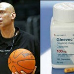 Kareem Abdul-Jabbar and the Magic Cancer Bullet comment by Kevin Li
 Kelly Ripa Diagnoses Herself With Mysterious Neurological Disorder comment by Kevin Li
 Why is Miley Cyrus Harming Herself? comment by Monica Bodd
 "Painfully Awkward Rob Lowe" Ad under Attack by "Shy Bladder" Advocates comment by Angelina Iyinbor
 Does Heidi Montag have a plastic surgery addiction? comment by Rachel Zimmerman
 Amanda Bynes Diagnosed as Bipolar comment by Monica Bodd
 Avicii Hospitalized, to Undergo Gall Bladder Surgery comment by Kevin Li
 Gina Rodriguez Surprised by Hashimoto's Diagnosis- UPDATED comment by Hannah Willey


4 Comments